O-Glycan Profiling Analysis
Creative Proteomics specializes in glycomics services, including detailed O-glycan profiling, protein glycosylation analysis, and structural characterization. Utilizing advanced techniques like mass spectrometry and liquid chromatography, Creative Proteomics delivers accurate and customized reports to support research, clinical diagnostics, and biopharmaceutical applications, offering valuable insights into molecular structures and functions.
Submit Your Request Now
×
- Define
- What We Provide
- Technology Platform
- Advantages
- Sample Requirements
- Demo
- FAQs
- Case
- Publications
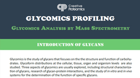
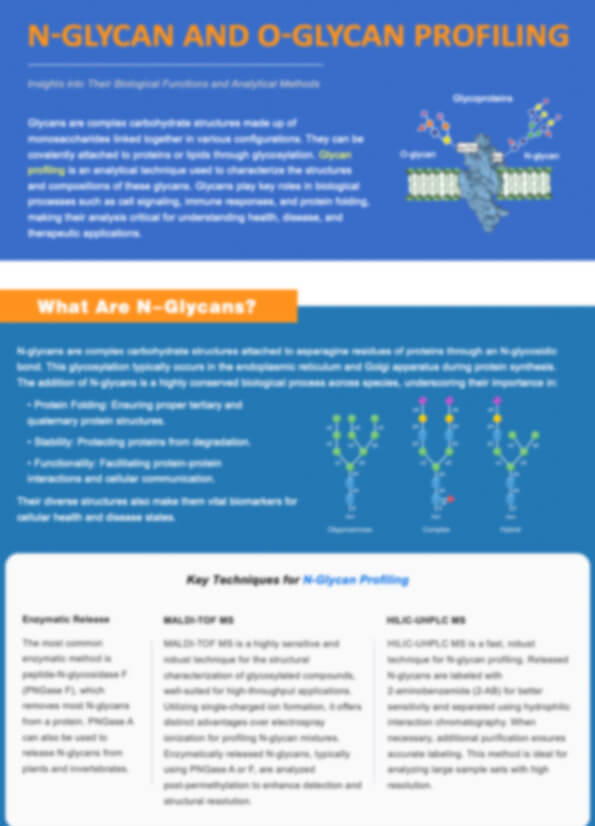
What are O-Glycans?
O-glycans are complex carbohydrate structures attached to proteins through a glycosidic bond between a monosaccharide and the hydroxyl group of serine or threonine residues. Unlike N-glycans, which are attached to asparagine, O-glycans exhibit significant structural heterogeneity, with varying chain lengths, branching patterns, and monosaccharide compositions. Commonly initiated by N-acetylgalactosamine (GalNAc), O-glycans can be classified into several families, including core 1 (Tn antigen) and core 2, each with distinct biological functions such as protein stability, immune response modulation, and cell signaling.
O-glycosylation plays a crucial role in various physiological processes, including cellular recognition, adhesion, and protection. The diversity and complexity of O-glycan structures make them challenging to study but essential in understanding diseases, particularly cancers, where altered glycosylation patterns are often observed. In the biopharmaceutical industry, O-glycan profiling is critical for optimizing therapeutic glycoproteins, as glycosylation directly influences their pharmacokinetics, stability, and immune interactions.
O-Glycan Profiling at Creative Proteomics
At Creative Proteomics, our O-glycan profiling service includes comprehensive analysis using advanced techniques to identify and characterize the various O-glycan structures present on proteins. Our service provides data that is essential for both basic research and applied industries, such as biopharmaceuticals and diagnostics.
Release and Isolation
O-linked glycans are typically released from glycoproteins through reductive alkaline β-elimination. The released O-glycans are then isolated and enriched using hydrophilic interaction chromatography (HILIC), which helps to separate glycans based on their hydrophilicity.
Exoglycosidase Digestion
To determine the linkage and structure of each glycan form, we perform specific exoglycosidase digestion. This step allows for detailed characterization of the glycosidic linkages, providing crucial information about the glycan's composition.
Labeling for Enhanced Detection
For enhanced sensitivity in high-performance liquid chromatography (HPLC) and mass spectrometry (MS)-based analyses, O-glycans are often fluorescently labeled using reagents such as 2-aminobenzoic acid (2-AA) or 2-aminobenzamide (2-AB). These labeling techniques enable more precise detection and analysis of glycan profiles.
Advanced Analytical Techniques
The labeled and enriched O-glycans are then analyzed using mass spectrometry techniques, including MALDI-TOF MS and ESI-MS. These methods provide detailed information on the glycan structures, including their molecular weight, composition, and branching patterns.
Comprehensive Data Analysis
Our experts perform a detailed analysis of the mass spectrometry data, providing high-resolution insights into the composition of the O-glycans present. The results are delivered in a comprehensive report, which includes the identification of glycan structures and detailed analysis of their molecular weights and glycosidic linkages.
 The general procedure of sample preparation for glycan profiling
Technology Platforms for O-Glycan Profiling
Matrix-Assisted Laser Desorption/Ionization Time-of-Flight Mass Spectrometer
MALDI-TOF MS is employed to analyze the permethylated O-glycans in the positive ion mode. This technique offers high sensitivity and resolution, allowing for the detection of various glycan isoforms and their detailed structural features.
Electrospray Ionization Mass Spectrometer
ESI-MS provides high accuracy in determining the molecular weight and composition of glycans in solution. It is particularly useful for identifying glycan structures and the linkages between monosaccharides.
High-Performance Liquid Chromatography
HPLC, coupled with fluorescent labeling reagents like 2-AA or 2-AB, is used for the separation and quantification of released O-glycans. This method enhances the resolution of complex glycan mixtures before MS analysis, ensuring accurate profiling of glycan structures.
Advantages of O-Glycan Profiling Service
- Comprehensive Structural Analysis: Detect and characterize over 50 different O-glycan structures, including variations in branching and monosaccharide composition.
- High Throughput: Process up to 100 samples per week, making it ideal for large-scale studies and biopharmaceutical applications.
- High Sensitivity: Achieve detection of glycan structures at concentrations as low as 1 ng using advanced MALDI-TOF MS and ESI-MS techniques.
- Accurate Quantification: Provide quantitative data with over 90% accuracy, ensuring precise comparisons of glycosylation patterns.
- Tailored Reporting: Deliver customized reports with focused data interpretation, supporting research and therapeutic glycoprotein development.
Sample Requirements for O-Glycan Profiling
| Sample Type | Required Amount | Format | Condition | Additional Notes |
|---|---|---|---|---|
| Glycoprotein Sample | 1-10 mg (protein concentration) | Liquid or Lyophilized | -20°C or -80°C (Frozen) | Ensure the sample is free of contaminants, such as salts or detergents. |
| Peptide Sample | 50-100 µg | Liquid or Lyophilized | -20°C or -80°C (Frozen) | Avoid protease activity during storage. |
| Cell Extract | 1-2 mL | Liquid | -80°C (Frozen) | Preferably fresh or snap-frozen. |
| Tissue Sample | 10-50 mg | Fresh or Frozen | -80°C (Frozen) | Samples should be homogenized before submission. |
| Plasma/Serum | 200 µL - 1 mL | Liquid | -80°C (Frozen) | Must be centrifuged to remove any cellular components. |
| Urine/Saliva | 500 µL - 1 mL | Liquid | -80°C (Frozen) | Collect in sterile containers to avoid contamination. |
Demo

Example of O-Glycan structures detected and analyzed by using MS (S. Yang et al., 2017)
FAQ of O-Glycan Profiling
What is the ideal sample preparation for O-glycan profiling?
The ideal sample for O-glycan profiling should be free of contaminants such as salts, detergents, and high concentrations of small molecules that could interfere with the analysis. For glycoprotein samples, we recommend isolating the protein of interest using standard purification techniques before sending it for profiling. In the case of peptide samples, ensure they are free from proteolytic activity, which can degrade the O-glycans. For cell or tissue extracts, ensure that the samples are homogenized and stored at -80°C to preserve glycan structures.
How should I store my samples before submission?
Samples should be stored at -80°C to ensure the integrity of O-glycans. Freezing at this temperature prevents enzymatic degradation and maintains the structure of the glycans. If shipping is required, samples should be shipped on dry ice to prevent thawing during transit. For lyophilized samples, ensure they are tightly sealed in moisture-proof containers.
What types of samples can be analyzed for O-glycans?
We accept a wide range of biological samples, including purified glycoproteins, peptides, plasma, serum, cell extracts, and even tissue samples. It is important to ensure that the sample is appropriate for O-glycan analysis—glycoproteins are the most commonly analyzed, as they are directly linked to O-glycosylation. Additionally, for tissue or cell extracts, efficient extraction and proper sample preparation are crucial for optimal results.
How long does the O-glycan profiling analysis take?
The typical turnaround time for O-glycan profiling is around 2-4 weeks, depending on the complexity and number of samples. More complex samples with a higher degree of glycosylation may require additional time for detailed analysis. We provide progress updates throughout the process and ensure timely delivery of final reports.
Can you analyze low-abundance O-glycans in complex samples?
Yes, our advanced mass spectrometry techniques, including MALDI-TOF MS and ESI-MS, offer high sensitivity and can detect low-abundance O-glycans at concentrations as low as 1 ng. The use of fluorescent labeling and enrichment strategies further improves the detection of trace amounts of O-glycans in complex samples, ensuring that even subtle glycosylation changes are identified.
What if my sample contains mixed glycan types?
Our analytical methods are highly versatile and can distinguish between different glycan types, even in complex mixtures. We can separate and identify multiple O-glycan isoforms through our combination of chromatographic and mass spectrometric techniques. Additionally, we can provide detailed structural characterization of each glycan present in the sample, ensuring you get a comprehensive analysis.
Learn about other Q&A.
O-Glycan Profiling Case Study

Publications
Here are some publications in proteomics research from our clients:

- Conformational differences between functional human immunodeficiency virus envelope glycoprotein trimers and stabilized soluble trimers. 2019. https://doi.org/10.1128/jvi.01709-18
- Identification of the O-Glycan Epitope Targeted by the Anti-Human Carcinoma Monoclonal Antibody (mAb) NEO-201. 2022. https://doi.org/10.3390/cancers14204999
- Alternative glycosylation controls endoplasmic reticulum dynamics and tubular extension in mammalian cells. 2021. https://doi.org/10.1126/sciadv.abe8349
- Modulation of the Endomembrane System by the Anticancer Natural Product Superstolide/ZJ-101. 2023. https://doi.org/10.3390/ijms24119575